Pediatric Cardiac Surgery Lab
The Stanford Pediatric Cardiac Surgery Lab research program is focused on three aspects of surgery for congenital cardiac disease:
We are developing surgical and technological approaches to permit the repair of heart defects in utero.
We would like to make it possible to surgically correct congenital heart defects in utero. Such early intervention could significantly reduce morbidity because once abnormal blood flow patterns begin, they prevent the normal structural development of the heart, requiring complex surgeries after birth.
We want to make pediatric cardiac surgery as safe as possible.
We are working to develop surgical approaches to protect the developing neonatal brain from injury during complex surgeries that require use of the heart-lung machine.
We are developing novel strategies for bioengineering heart valves for pediatric patients.
We are working to bioengineer pediatric heart valves that can self-renew and grow with the child.

Our open lab positions are listed below. If you are interested in our research or lab-related research opportunities, please contact:
R. Kirk Riemer, PhD
Research Lab Director
Phone: (650) 723-8958
riemerk@stanford.edu
Cardiac Bypass in the Fetus
We believe that surgical correction of certain congenital heart defects in utero is feasible and provides the opportunity for significant reduction in morbidity by restoring normal blood flow patterns through the heart and great vessels during development. It has been shown that the bulk of the cardiac maldevelopment is secondary to the disturbed flow patterns that arise, for example, when the tricuspid valve fails to develop: During fetal life in the presence of tricuspid atresia, the failure of blood to flow into the right ventricle (RV) causes hypoplasia of the RV. If the normal flow pattern through the valve could be re-established early in gestation, the flow-dependent pathophysiology should be prevented. This is the rationale for fetal cardiac surgery.
To perform surgery on intracardiac structures or great vessels, it is necessary to put the fetus on extracorporeal circulation (i.e., cardiac bypass using a heart-lung machine). Since the fetus is effectively already on physiologic “bypass” via the placenta, the process is much more complex than it is after birth and presents many unique challenges. Our ongoing studies show that cardiac bypass can be safely performed in fetuses as small as 450 grams without transfusion. We have demonstrated that several critical variables — including anesthesia modality, fetal temperature changes, umbilical cord manipulation, uterine tone, and maternal blood pressure — interact in complex ways that can result in compromised utero-placental gas exchange following the return from bypass.
These fetal surgical intervention studies have been supported in part by the Oak Foundation.
Cerebral Protection During Cardiopulmonary Bypass
Protecting the brain during heart surgery using regional low-flow perfusion (RLFP) during deep hypothermic circulatory arrest (DHCA) is critical for pediatric patients because their developing brains are particularly vulnerable to hypoxic damage. Such damage may occur during the extended periods of circulatory arrest needed for repair of complex congenital cardiac lesions. We have recently begun to model the protective effects of RLFP during DHCA on neuronal and glial cell apoptosis in neonatal animal models. These studies seek objective evidence of the beneficial effects of RLFP on brain apoptosis. We are in the process of analyzing the data from a recent series of such studies that we expect will demonstrate the ability of RLFP to reduce cellular apoptosis in vulnerable brain regions. These studies have now been are presently extended to include evaluation of neuronal damage via MRI.
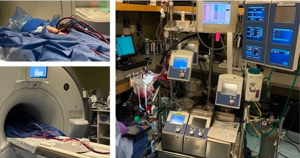
A major externally funded project in the Peds Cardiac Division is using Magnetic Resonance Spectroscopy to measure metabolites in the brain during different modes of cardiopulmonary bypass in neonatal piglets.
Tissue Engineering for Pediatric Applications
Our Lab has recently launched a program in heart-valve tissue biology and bioengineering to meet the unique needs of pediatric patients.
Because a child’s heart and great vessels are still growing rapidly, children often require additional surgeries when they outgrow the graft materials currently available for congenital heart defect repairs. Because these non-growing tissues patches also do not repair themselves, they often fail too early in pediatric patients. We are developing the technology to produce transplantable tissues that can grow with children.
The efforts of the Lab are initially focused on bioengineering pediatric heart valves. Experiments are underway to discover and decode the mechanisms through which valves self-repair, using a novel culture system that replicates the biomechanical stimuli experienced by heart valves in vivo. This area of fundamental biology is critical to the goals of bioengineering custom-sized replacement tissues for children’s hearts.
We are collaborating with scientists in medical as well as engineering disciplines at Stanford in a team approach to this project. We believe our research will support complementary methods now being tried around the world to generate engineered tissues. Our heart valve regenerative biology studies are largely underwritten by the Alex Vibber Endowment and also supported by the Oak Foundation.