Marsupials have short pregnancies. Their placentas mimic those of mice during early fetal development, while other key placental genes are expressed and secreted into milk for the offspring, Stanford researchers say.
September 12, 2017 - By Krista Conger
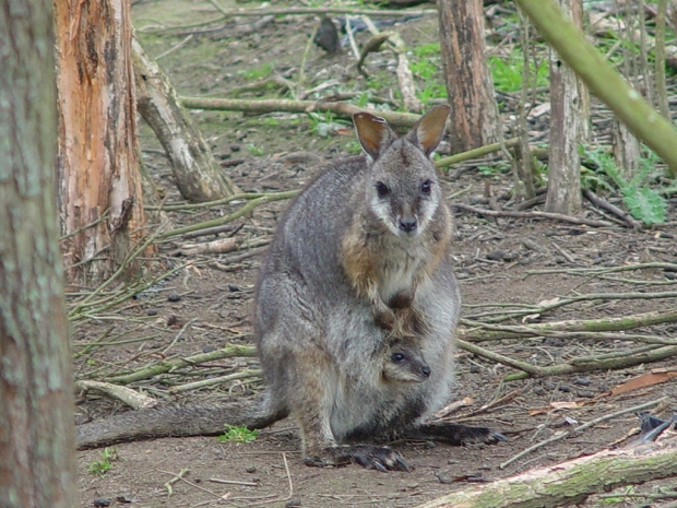
Researchers studying the tammar wallaby discovered that the female marsupials express genes important for fetal development in their milk.
MB Renfree
Modern mothers, whether they be human or mouse, might be forgiven for envying marsupial mamas. Rather than enduring a long pregnancy and the birth of a relatively well-developed — and comparatively large — baby, kangaroos, wallabies and their ilk blithely pop out offspring after pregnancies measured in days rather than months.
These tiny, almost formless creatures then make their own intrepid way up to the mother’s pouch to nestle politely and nurse for sometimes as long as a year.
For decades, researchers assumed that this premature eviction from the womb left little or no role for the placenta, which in most mammals tightly links the physiological processes of the mother and the fetus to support the fetus’s many stages of development. These mammals are called eutherian mammals to distinguish them from the evolutionarily distant marsupials. In the past decade or so, however, it has become apparent that marsupials do sport their own, rudimentary version of this important organ.
Now researchers at the Stanford University School of Medicine and the University of Melbourne in Australia have collaborated to learn that marsupials have evolved a clever trick to support their need for a shortened pregnancy and a long lactation period. In short, female marsupials express genes important for fetal development that are normally found in the later stages of the eutherian placenta in their mammary glands instead — a kind of handoff of the developmental baton from womb to milk that suits their unique, savanna-hopping lifestyle.
“This research basically shows that the placenta, while really different-looking in the marsupial, has many of the functions of the eutherian,” said Julie Baker, PhD, professor of genetics at Stanford. “Each animal has come up with their own unique strategies for delivering the functions of the placenta that takes into account where they live, how many offspring they have and what they eat, for example. But the actual function is very well-conserved.”
Baker shares senior authorship of the study, which was published online Sept. 12 in eLife, with Marilyn Renfree, PhD, a professor of zoology at the University of Melbourne. The lead author is Stanford graduate student Michael Guernsey.
A little wallaby
Guernsey and Baker studied the placenta of the tammar wallaby, which is native to Australia. To the marsupially naïve, it resembles a tiny kangaroo. Males weigh no more than 20 pounds and stand about 18 inches high. It forages hoppily by night. The tammar wallaby has a pregnancy that lasts a mere 26.5 days, after which the young climb into the pouch and nurse for the next 300 to 350 days as they complete their development.
The wallaby’s placenta is deceptively simple.

Julie Baker
“There are only two main tissue types,” said Guernsey, “one responsible for nutrient distribution and one for respiration. We wanted to see which, if any, gene products found in the eutherian placenta are also in the marsupial placenta, and where they are expressed. Conversely, which eutherian markers might be missing?”
In contrast, the eutherian placenta is highly complex and comprises both maternal and fetal tissue.
Guernsey studied the RNA transcripts in the wallaby placenta and compared them with those found in eutherian mammals during various stages of fetal development. He found that the gene expression patterns in the marsupial placenta undergo dynamic, rapid changes during the last few days of the animals’ short pregnancy, during which the placenta churns out proteins known to be important in the early stages of eutherian development.
“All of the wallabies’ gene expression time points were most similar to those found in the early eutherian placenta,” said Guernsey. “But where have the late functions of the eutherian placenta gone?”
Changes in the milk
A key might lie in the complex makeup of the animals’ milk, the composition of which changes to meet the demands of the growing, pouch-bound youngster. It’s so potent that placing an infant into the pouch of a mother who has been nursing a more developmentally enhanced baby causes the newcomer to beef up dramatically, increasing its head size and body weight and growing thicker fur than its age-matched peers.
To investigate the relationship between the marsupial placenta and the milk produced during lactation, the researchers homed in on 77 genes whose expression was shared among the tammar placenta, the eutherian placenta and the tammar mammary gland, but not the mouse mammary gland.
Essentially, we’re trying to understand how the placenta evolved in the first place.
Many of the genes they identified were associated with nutrient transport. Another, known as GCM1, is a transcription factor essential to the function of eutherian placentas.
“This is the first documented expression of GCM1 outside the placenta in mammals,” said Baker. “What we’re learning is that the marsupial placenta functions much as it does in eutherians in the very early stages of development, but the expression of later-stage eutherian placental genes instead occurs in the mammary gland. So clearly although the placentas of humans, cows or mice are extraordinarily different from those of marsupials, the animals are fulfilling the same necessary functions in different ways.”
“Essentially, we’re trying to understand how the placenta evolved in the first place,” said Guernsey. “It’s a difficult question to answer. We’re finding that we can begin to identify rudimentary placentas in other species as well, like lizards and fish. It will be really interesting to see whether, in this completely different evolutionary landscape, these functions are still conserved in ways that make sense for that animal.”
Baker is a member of Stanford Bio-X and the Stanford Child Health Research Institute.
Guillaume Cornelis, PhD, a postdoctoral scholar at Stanford, is also a co-author of the study.
The research was supported by a National Science Foundation, the Berry Foundation and the Australian Research Council.
Stanford’s Department of Genetics also supported the work.
-
 Krista CongerKrista Conger is a senior science writer in the Office of Communications. Email her at kristac@stanford.edu.
About Stanford Medicine
Stanford Medicine is an integrated academic health system comprising the Stanford School of Medicine and adult and pediatric health care delivery systems. Together, they harness the full potential of biomedicine through collaborative research, education and clinical care for patients. For more information, please visit med.stanford.edu.

