Ongoing Research
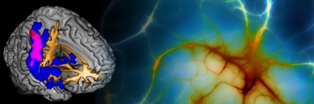
Dr. Poston's research interests include the development, validation, and application of functional and structural neuroimaging as biomarkers for the diagnosis and treatment of movement disorders. Specifically, her research focuses on using FDG PET, functional MRI (fMRI), and ultra-high resolution structural MRI (7T MRI) to understand abnormal brain networks that lead to both motor and cognitive dysfunction in patients with Parkinson's disease, multiple system atrophy, progressive supranuclear palsy, and corticobasal syndrome/corticobasal degeneration.
Recruiting Now
Mind & Memory Changes Study (LB-SPARK)
The Lewy Body Scientific Partnership for Advancing Research and Knowledge aims to understand mind and memory changes in Parkinson’s disease. LB-SPARK is recruiting individuals with Parkinson’s disease, Dementia with Lewy bodies, and healthy volunteers.
This is a longitudinal study looking at Parkinson’s disease over time. You will be contacted once per year by phone or visit at Stanford. Study participation includes neurological exams, neuropsychology testing, a blood sample, gait & balance tests, collecting cerebrospinal fluid (optional), and collecting PET/MRI scans (optional).
Contact
For more information, contact: Hannah Schmitz
If you are interested in participating in LB-SPARK, please fill out this intake form and a member of the study team will contact you.
Facial Expression Biomarker Study
We are looking for volunteers to participate in a study looking at facial movements in neurodegenerative diseases. We hope to validate a diagnostic tool developed by Stanford Undergraduate, Erin Smith, for early detection of Parkinson’s disease. Study participants will be asked to come in for one 30 minute study visit.
Subjects
We are recruiting patients with Dementia with Lewy bodies, Mild Cognitive Impairment due to Lewy bodies, Parkinson’s disease, REM Sleep Behavior Disorder, Multiple System Atrophy, Progressive Supranuclear Palsy, Corticobasal Syndrome, and Healthy Control participants, ages 18 years or older.
Contact
If you are interested in learning more about participation, please contact Alena Smith at alenaa@stanford.edu or (650) 723-0060.
PET Study
We recently started recruiting individuals with Parkinson’s disease for two PET/MRI scans to examine patterns of protein (amyloid) build up and inflammation (microglial activation) in the brain. The ability to measure amyloid accumulation and microglial activation provides a critical opportunity to further the understanding of Parkinson’s disease, monitor disease progression, and assess disease modifying treatments.
Subjects
People with Parkinson’s disease over age 50, with and without cognitive or memory problems.
Contact
If you are interested in learning more about participation, please contact Marian Shahid by email at mshahid@stanford.edu or at (650) 723-0060.
Healthy Brain Aging Study
Our goal for the research is to follow volunteers over time, including eventual brain donation, to develop biological markers that enhance understanding, early detection and effective treatment of Parkinson’s disease, Lewy body disease, Alzheimer’s disease, age-related cognitive decline and similar disorders of the aging brain. It is funded by the National Institute of Health and is part of the Alzheimer’s Disease Research Center (ADRC).
Subjects
Parkinson’s disease or Lewy body disease, without memory problems or with mild-to moderate problems, those with Alzheimer’s or mild cognitive impairment as well as healthy controls.
Contact
If you are interested in learning more about participation, please contact Veronica Ramirez or Christina Wyss-Coray
Stanford Brain Bank Program
This study seeks a broad range of participants with neurological disorders, including Alzheimer’s disease, Parkinson’s disease, Lewy Body disease, and other disorders of the brain. We also seek healthy older adults age 65 and older without a brain disorder to serve as controls. Participants will be asked to consider brain donation at the time of death. Autopsy tissues will be used to help our scientists better understand neurodegenerative disorders and other brain diseases, by comparing clinical and autopsy findings and by examining microscopic, biochemical and genetic features. The pathologist’s brain autopsy report will be provided to family members.
PI: Victor Henderson, MD, MS, Co-PI: Edward Plowey, MD, PhD
Subjects
Open, enrollment ongoing, limited to current or past Stanford Health Care patients
Contact
If you are interested in learning more about participation, please contact Christina Wyss-Coray, RN, BSN, PHN, by email at ADRCstanford@stanford.edu, or at (650)721-2409.
Ongoing, Closed to recruitment
Development of Multimodal Imaging Biomarkers for Cognitive Dysfunction in Parkinson’s Disease
- Why do some people with Parkinson’s disease develop memory problems soon after diagnosis, but for others it takes many years?
- Why do some people with Parkinson’s disease develop dementia, but for others memory problems are very mild?
- How common is it for people with Parkinson’s disease to also develop Alzheimer’s disease?
The goal of this study, funded by the Michael J Fox Foundation for Parkinson’s disease Research (MJFF), is to help answer these questions about cognition and PD. This study is part of the MJFF initiative “Defining Cognitive Phenotypes of Parkinson’s Disease 2011”. For this study, participants take part in MRI scans, as well as clinical evaluations of movement and memory. Participants also undergo a blood draw and a lumbar puncture (FAQs). We are currently enrolling people with Parkinson’s disease who do not have memory problems, people with Parkinson’s disease who do have memory problems, and non-Parkinson’s disease (control) participants who do not have memory problems.
Early Differential Diagnosis of Parkinsonism with Metabolic Imaging and Pattern Analysis
- Can functional neuroimaging improve the diagnostic accuracy for Parkinson’s disease and other parkinsonian disorders (such as Multiple System Atrophy and Progressive Supranuclear Palsy)?
In 2010 we published a proof-of-principle study that suggests a functional neuroimaging technique called 18F-fluorodeoxyglucose (FDG) positron emission tomography (PET) can help diagnose people with Parkinson’s disease, Multiple System Atrophy, and Progressive Supranuclear Palsy (read paper). This large, multi-center study aims to determine how sensitive and specific this imaging technique is for accurate diagnosis in people who are just developing symptoms. In addition, we aim to determine if this technique can be used to predict if persons at risk for developing parkinsonism, such as persons with REM sleep Behavior Disorder (RBD), will indeed develop Parkinson’s disease or Multiple System Atrophy. We are currently recruiting people with Parkinson’s disease, Multiple System Atrophy, Progressive Supranuclear Palsy, and Corticobasal Syndrome (CBS). We are also recruiting people who have just recently developed parkinsonism, even if the diagnosis is uncertain. Finally, we are recruiting people who have RBD, but do not currently have any symptoms of parkinsonism. This study is funded by the NIH/NINDS.
A 12-Week, Multicenter, Randomized, Parallel-Group Study to Assess the Safety, Tolerability, Pharmacokinetics, Biomarker Effects, Efficacy, and Effect on Microglia Activation, as Measured by Positron Emission Tomography, of AZD3241 in Subjects with Multiple System Atrophy
This multicenter study will enroll 64 patients with Multiple System Atrophy. The objectives of this study are to assess the safety, tolerability, efficacy, and effect on microglia activation (as measured by PET) of two dosage levels of AZD3241 versus placebo (non-active study drug) in subjects with MSA. AZD3241 is an investigational drug, not approved by the FDA. The total length of the study is approximately 16 weeks, with 12 weeks of study medication.
For more information, please visit: https://clinicaltrials.gov/ct2/show/NCT02388295
Attention and Working Memory in Parkinson's Disease
- Do dopamine medications improve or worsen Attention and Working Memory in people with Parkinson’s disease?
Currently, researchers do not understand how dopamine influences aspects of cognition, such as the ability to pay attention to what is important, and the ability to keep in mind new information once it is learned. This study aims to answer this question. Participants are tested on a task of Working Memory (the ability to quickly remember new information) and Attention (the ability to focus on what is important) while both ON and OFF of dopamine medications.
A Phase 1/2 Trial Assessing the Safety and Efficacy of Bilateral Intraputaminal and Intranigral Administration of CERE-120 (Adeno-Associated Virus Serotype 2 [AAV2]-Neurturin [NTN]) in Subjects With Idiopathic Parkinson’s Disease
- Does gene-therapy of AAV2-NTN help slow the progression of Parkinson’s disease motor symptoms?
While this treatment appeared to be safe, the 15 month follow up data showed that there was no difference in the progression of clinical motor symptoms between patients who received the therapy and patients who received a ‘sham’ surgery (i.e. did not receive the therapy). The long-term follow up of these patients is currently underway.
Basal Ganglia Modulation of Cognitive Systems in Parkinson’s Disease
- How do dopamine replacement medications (such as ropinerole, pramipexole, and levodopa) change cognition in people with Parkinson’s disease?
Unfortunately, medications to help the motor symptoms of Parkinson’s disease, such as levodopa, do not usually improve memory problems sometime encountered by patients. However, these medications can improve some aspects of thinking; but they also have the potential to worsen other aspects of thinking. The purpose of this study is to use functional neuroimaging to answer these questions. This study has completed its first phase, which is currently being analyzed (results to be given on the Research Alumni page). This study is funded by the NIH/NINDS.
Structural Correlates of Cognitive and Motor Dysfunction in Parkinsonian Disorders
- Can we use ultra-high resolution MRI scanning to help diagnose patients with Parkinson’s disease?
- Can we detect small changes in brain structures that are associated with specific motor symptoms and cognitive symptoms of Parkinson’s disease?
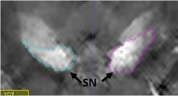
At Stanford we use an exceptionally high-powered MRI magnet (called a 7-Tesla MRI) to look at tiny structures in the brain. This MRI scanner can show details of the brain down to 200 μm, or one-fifth of a millimeter. We are able to produce ultra-high resolution images of the Substantia Nigra (SN) and the Subthalamic nucleus (STN) (see images below), two very important structures in Parkinson’s disease pathology. We are also able to view the hippocampus, a critical brain region involved with memory (paper).

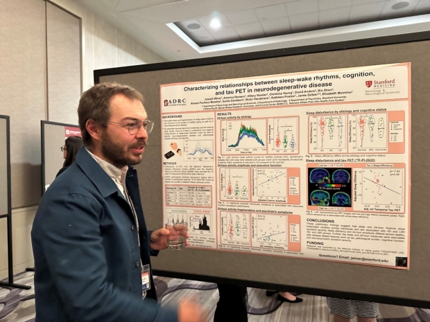
Dr. Joe Winer presenting research at the Spring 2023 Alzheimer’s Disease Research Center (ADRC) Meeting in Washington, D.C.

Dr. Carla Abdelnour presenting at the 2023 Alzheimer’s & Parkinson’s Diseases Conference (ADPD) in Gothenburg, Sweden.
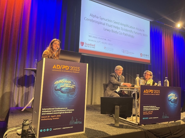
Dr. Melanie Plastini presenting at the 2023 Alzheimer’s & Parkinson’s Diseases Conference (ADPD) in Gothenburg, Sweden.
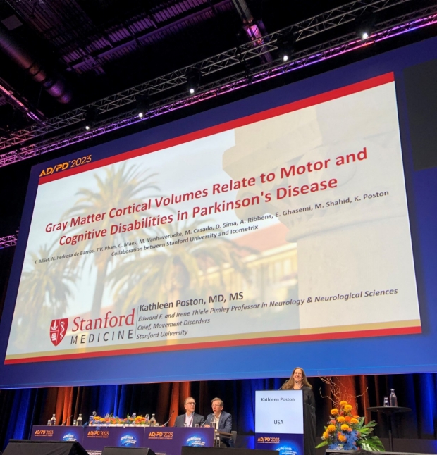
Dr. Kathleen Poston presenting at the 2023 Alzheimer’s & Parkinson’s Diseases Conference (ADPD) in Gothenburg, Sweden.

Dr. Carla Abdelnour presenting at the 2022 Clinical Trials on Alzheimer’s Disease (CTAD)
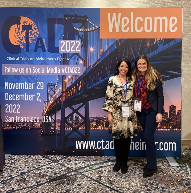
Carla (left) and Melanie (right) at the 2022 Clinical Trials on Alzheimer’s Disease (CTAD)
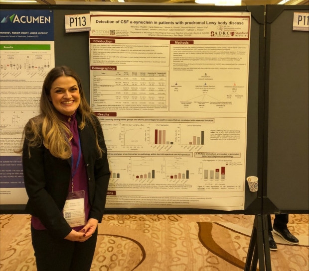
Dr. Melanie Plastini presenting at the 2022 Clinical Trials on Alzheimer’s Disease (CTAD)
Alena (left) and Carla (right) at the 2022 Wu Tsai Neuroscience Symposium

Dr. Carla Abdelnour presenting at the 2022 Wu Tsai Neuroscience Symposium
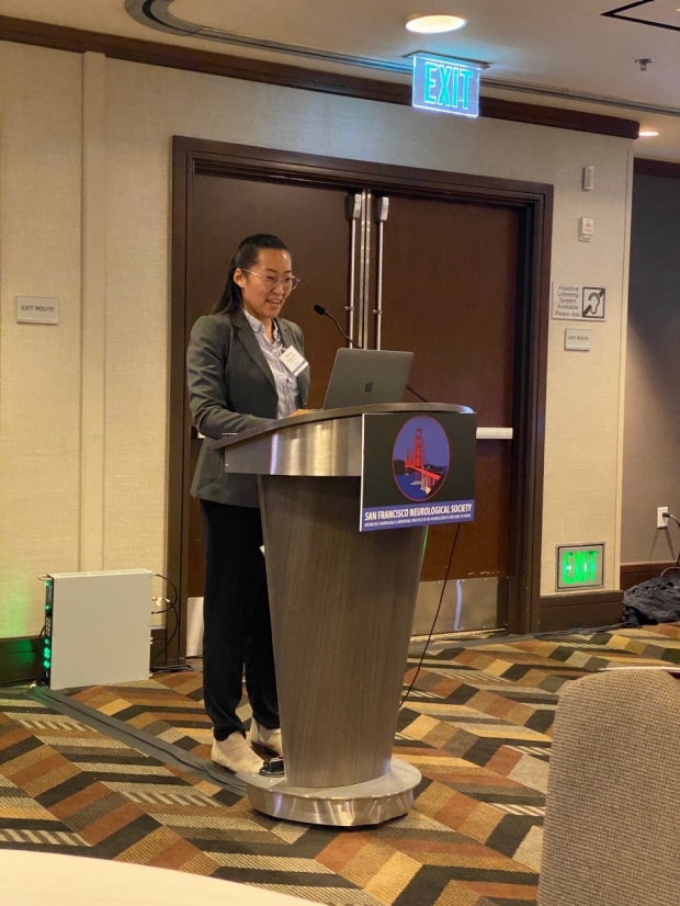
Dr. Christina Young presenting at the 2022 San Francisco Neurological Society (SFNS)

Marian presenting at the 2022 International Congress of Parkinson’s Disease and Movement Disorders in Madrid, Spain

Dr. Carla Abdelnour presenting at the 2022 International Congress of Parkinson’s Disease and Movement Disorders in Madrid, Spain
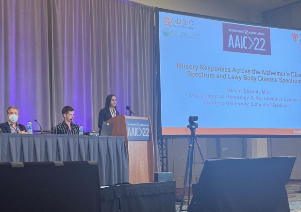
Marian presenting at the 2022 Alzheimer’s Association International Conference (AAIC) in San Diego, CA

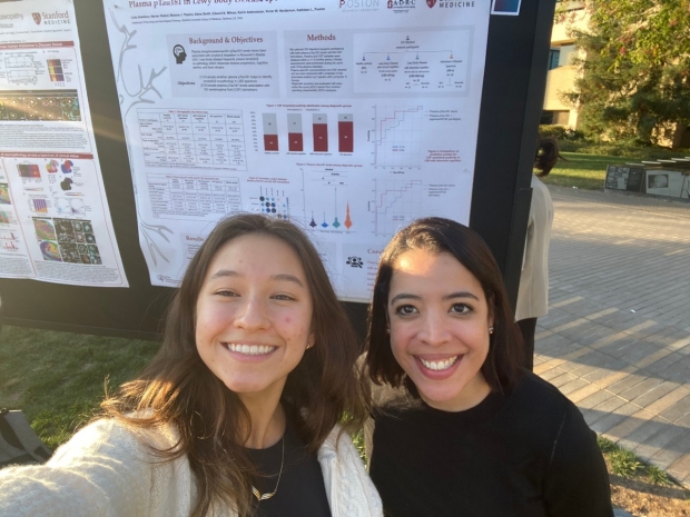
Dr. Christina Young presenting at the 2022 Alzheimer’s Association International Conference (AAIC) in San Diego, CA

Dr. Poston & Jee Kim at the 2019 American Neurological Association (ANA) Annual Meeting

Dr. Kathleen Poston at AAN TRANSCENDS mentee Dr. Chantale Branson’s research presentation at the 2019 AAN
AAN TRANSCENDS Scholars program: Mentors Dr. Kathleen Poston and Dr. Michael Schwarzschild with Mentee Dr. Chantale Branson.

Colin and Nessa presenting at the 2019 Cognitive Neuroscience Society Annual Meeting
Veronica Ramirez presenting at the 2018 American Academy of Neurology Annual Meeting

Dr. Zhang presenting at the 2017 Organization for Human Brain Mapping Annual Meeting

Dr. Poston and Dr. Linortner at the NIH/NINDS Udall Centers Directors' Meeting in Washington DC