Stanford Pituitary Center - Surgical Approach

Our surgical team, led by Dr. Fernandez-Miranda, provides patients the latest surgical techniques for pituitary tumor removal and remission. Dr. Fernandez-Miranda is an internationally renowned surgical innovator and pioneer in endoscopic endonasal surgery for complex pituitary tumors. He has performed more than 1000 endoscopic endonasal operations, many of very high complexity, representing one of the largest endoscopic surgical series to date.
Dr. Fernandez-Miranda is recognized worldwide as a master in surgical neuroanatomy, the basis for accurate, safe, and gentle surgery. He has developed new surgical techniques for pituitary tumors invading the cavernous sinus and extending into the brain space that allow for higher rates of complete tumor removal and long-term remission in functional tumors.
Cushing's Disease and Negative MRI
Patients with Cushing’s disease often present with a “negative” MRI study, showing no evidence of tumor. Inferior petrosal sinus sampling is mandatory to confirm that the source of ACTH production is the pituitary gland.
The first step is these cases is to make sure the MRI study is of high-quality, as frequently patients do not obtain the best possible imaging. At Stanford Pituitary Center we have developed with our expert neuroradiologists the best possible imaging protocol to maximize chances of microadenoma identification; this includes a 1-mm fine-cut T1 sequences with and without contrast in all 3 spatial planes, dynamic sequences, and a flair post-contrast sequence.
If imaging is still not conclusive, intraoperative exploration is required, following the technique described by Prof. Oldfield that Dr. JFM learnt from him during his tenure at University of Virginia (2007-2008). This starts with wide surgical exposure to carefully explore all surfaces of the gland, including those in contact with the medial wall of the cavernous sinus, as microadenomas may cause mild bulging in the surface of the gland. Next, vertical incisions separated by 2-mm are performed to gently explore the inside of the gland, starting at the site of highest suspicion. Finally, when no adenoma is identified and selective removal is not possible, a partial hypophysectomy is performed by removing a quarter of the gland on each side and at the bottom of the gland, and preserving the central core of the gland. This technique maximizes chances of microadenoma removal while minimizing risk of pituitary dysfunction.

Cavernous Sinus Invasion
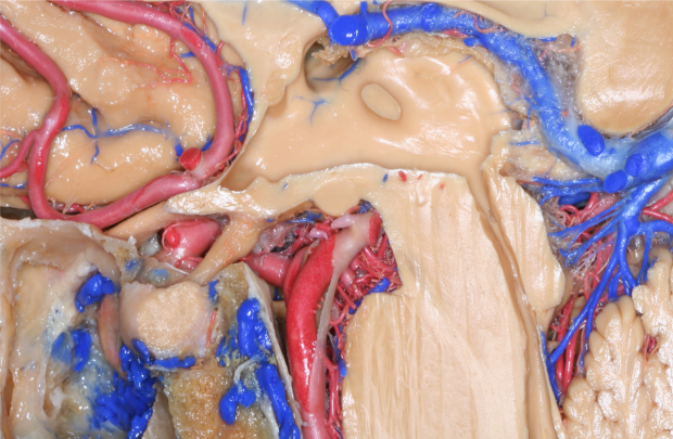
Pituitary tumors with cavernous sinus invasion represent a neurosurgical challenge. Safe and effective surgery in this area requires deep understanding of the cavernous sinus anatomy from an endonasal perspective.
Dr. JFM has been studying the cavernous sinus for over 15 years and has developed a surgical anatomy-based classification of the cavernous sinus with demonstrated utility for preoperative surgical planning and intraoperative guidance in pituitary surgery.
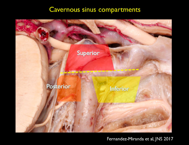
Four cavernous sinus compartments are described based on their spatial relationship with the cavernous carotid artery: superior, posterior, inferior, and lateral. Each compartment has distinct boundaries and dural and neurovascular relationships.
This essential knowledge has been successfully applied by Dr. JFM in over a hundred patients with invasive pituitary tumors. Selected pituitary adenomas with cavernous sinus invasion can now be completely removed at the Stanford Pituitary Center.
Journal of Neurosurgery Publication: Cavernous sinus compartments from the endoscopic endonasal approach: anatomical considerations and surgical relevance to adenoma surgery
Case Example:
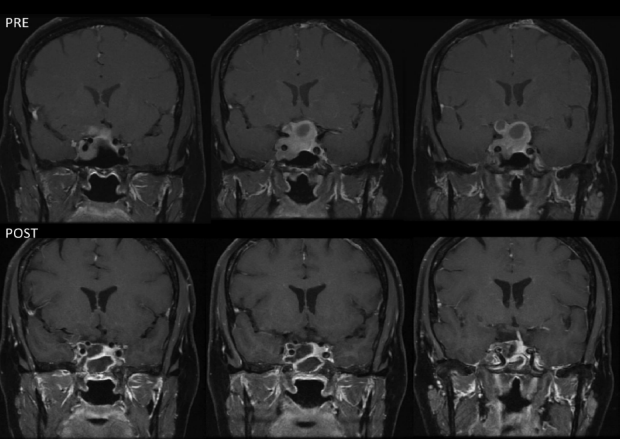
A 57 year-old women, presented with headaches and mild visual field defect. MRI study showed a large adenoma invading the superior and inferior compartments of the cavernous sinus, and compressing the optic apparatus. Careful preoperative planning and precise surgical technique allowed for complete tumor resection, with total recovery of preoperative deficits and no complications. The operative video can be reviewed here.
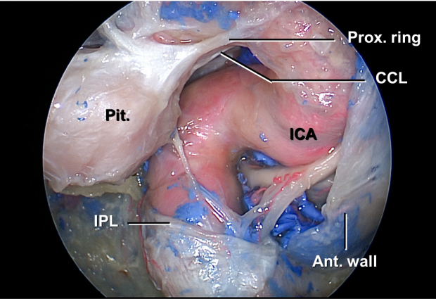
Selective Resection of the Medial Wall of the Cavernous Sinus
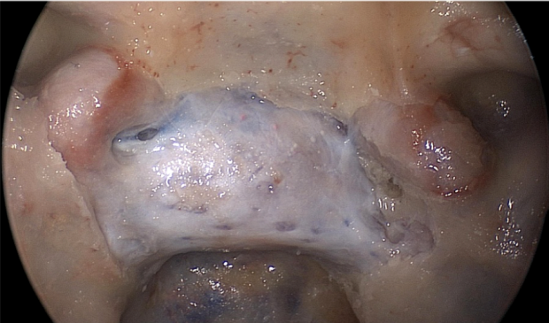
Pituitary adenomas often invade the medial wall of the cavernous sinus, but this structure is generally not surgically removed because of the risk of vascular and cranial nerve injury. This results on incomplete tumor resections and persistent disease in functional tumors.
Dr. JFM has meticulously investigated the medial wall of the cavernous sinus introducing a classification of the parasellar ligaments and their role in anchoring the medial wall, and has developed an innovative technique for selective resection of the medial wall when invaded by tumor.
At Stanford Pituitary Center, tumors invading the medial wall of the cavernous sinus can now be removed safely and effectively, with minimal morbidity and excellent resection and remission rates in hormonal-secreting adenomas causing Cushing's disease, Acromegaly, or Hyperprolactinemia.
Journal of Neurosurgery Publication: The medial wall of the cavernous sinus. Part 1: Surgical anatomy, ligaments, and surgical technique for its mobilization and/or resection.
Journal of Neurosurgery Publication: The medial wall of the cavernous sinus. Part 2: Selective medial wall resection in 50 pituitary adenoma patients.
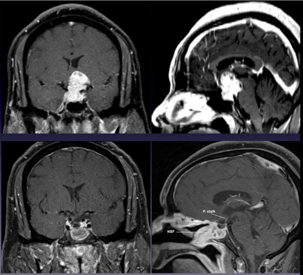
Case Example:
A 32 year-old man, presented with truncal obesity, diabetes, and hypertension; preoperative studies were compatible with Cushing’s disease. The MRI showed a large and invasive tumor. Intraoperatively it was invading the medial wall of the cavernous sinus bilaterally., and both were removed successfully achieving complete resection and functional remission that remains 5 years after surgery.


Transoculomor Triangle Approach for Adenomas Invading the Roof of the Cavernous Sinus
Pituitary adenomas may extend into the parapeduncular space by invading through the roof of the cavernous sinus. Currently, a transcranial approach is the preferred choice, with or without the combination of an endonasal approach.
Dr. JFM has described a novel surgical approach that takes advantage of the natural corridor provided by the tumor to further open the oculomotor triangle and resect tumor extension into the parapeduncular space.
The endoscopic endonasal transoculomotor approach is an original alternative for removal of tumor extension into the parapeduncular space in a single procedure.

Case Example:
A 62 year-old woman presented with headaches and episodes of double vision. Imaging studies showed a large adenoma with cavernous sinus invasion and extension into the parapeduncular space with compression of the cerebral peduncle and the medial temporal lobe. She was initially told that it was impossible to remove the parapeduncular extension of the tumor via endonasal endoscopic approach and will require a separate transcranial operation. However, a successful endoscopic endonasal surgery was performed with the recently described transoculomotor approach with near-complete resection, no complications, and without an additional craniotomy.

Complex Adenomas with Multiobular Shape and Subarachnoid Invasion
While most pituitary adenomas do not require extracapsular subarachnoidal dissection, there are complex adenomas with subarachnoidal invasion and multilobulated morphology, such as the one presented here, that require a combination of internal debulking, extracapsular and subarachnoidal dissection. The technique presented here allows for complete tumor resection, avoiding the risk of postoperative apoplexy of residual adenoma, and facilitates identification of perforating branches and neural structures that require meticulous preservation.
The patient is a 51-year-old male with profound visual loss with homonymous hemianopsia and left optic nerve atrophy. The options for surgical approach included transcranial, endoscopic endonasal, or a combination of both. An endoscopic endonasal approach was the preferred surgical option, because it allows for early identification of the pituitary gland, and provides access to the suprasellar region including pre- and retrochiasmatic spaces, which facilitates tumor removal while minimizing manipulation of the optic apparatus.
Watch videos of surgeries performed by Dr. Juan C. Fernandez Miranda. Warning: these videos contain surgical footage.

Featured Video
Stanford Pituitary Center